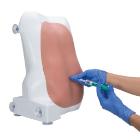
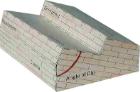
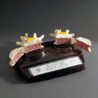
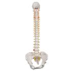
Epidural and Spinal Injection Trainer, Dimensions: 45 x 35 x 24 cm, functions: epidural anesthesia using the loss-of-resistance and the hanging-drop technique, spinal anesthesia with realistic resistance of the dura and arachnoid mater
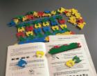
Replacement Standard LOR Kit (2) for epidural and spinal injection trainer, Replacement LOR kit, consisting of 2 loss-of-resistance inserts and 2 spinal tubes, long-lasting, very easy to change, Weight: 1.4 kg
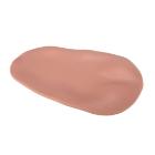
Replacement Geriatric LOR Insert for epidural and spinal injection trainer, to practice with the specific anatomical deformations of the spine of an elderly patient, easily replacable, Contents: Geriatric Insert, Spinal Tube
Replacement Geriatric LOR Kit (2) for Epidural and Spinal Injection Trainer, to practice with specific anatomical deformations of spine of an elderly patient, replacable standard patient LOR insert, Dimensions: 28x14x75cm
Horse Skull (Equus caballus), is a great way to study the anatomy of Equus caballus. This animal skull is perfect for mammal and comparative anatomy studies
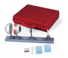
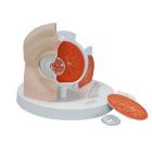
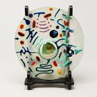
Model, Physical Eye, can be used to demonstrate the optical functions of the human eye, Half eyeball with adjustable iris diaphragm, lens holder and 2 convex lenses (f = 65 mm and 80mm), on a rod, Dimensions: 49 x 5.5 x 18 cm, Weight: 2.45 kg
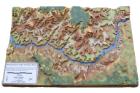
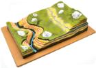
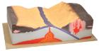
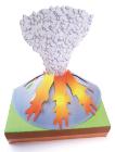
Tectonics Model, provide students with an innovative look at plate tectonics with this hand-painted plastic model, showing detailed representations of the earth layers. Both active and passive margins are shown, Oceanic floor spreading is also well-characterized
Additional SimScope* WiFi stethoscope, enables wireless communication between the Simscope* and the computer, allowing a seamless selection and change of pathological sounds and conditions of any standardised patient, O.S.C.E. Or low-fidelity manikin
SimScope WiFi* The Hybrid Simulator, enables wireless communication between the Simscope* and the computer, allowing a seamless selection and change of pathological sounds and conditions of any standardised patient, O.S.C.E. Or low-fidelity manikin
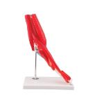
Model Elbow Joint 8 Parts, great tool for student and patient education, part of a high quality series of muscle models, and has been manufactured to replicate the anatomy of the human elbow joint in detail, colors used, Weight: 1.74 kg, Dimensions : 25 x 41 x 25 cm
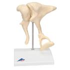
Model Ossicles Magnified 20 Times, joined to each other in the human body are located in the middle ear and are referred to as the auditory ossicles: malleus (hammer), incus (anvil) und stapes (stirrup), Weight: 0.385 kg, Dimensions : 17 x 12 x 21 cm